Student Guest Post: James Frazier on an Exploration of Taphonomy

During the fall semester (Sept-Dec 2018), students from Bryn Mawr and Haverford helped to clean and catalog the remains at Rutgers-Camden as part of a praxis course led by Professor Maja Šešelj. The following post if from one such student --- Bryn Mawr student James Frazier. James is a senior at Bryn Mawr College who is majoring in anthropology with a thesis on the taphonomy of the Arch St. remains.
Mystery Marks: An Exploration of Taphonomy
By James Frazier, Bryn Mawr College
A lot happens after bones make it into the ground, and the Arch St. Project creates a wonderful opportunity to see just what can happen. Taphonomy is the study of all the processes that affect a specimen, in this case human remains, from deposition to discovery and disinterment. There is a certain amount of damage inherent from being in the ground for 200-odd years, however each place where a specimen has been deposited, whether a cemetery or an open-air site, has unique factors to be considered. While existing research can provide broad expectations regarding the amount of damage that can be expected over time, each site is unique as the rate of taphonomic damage is influenced by burial practices, soil quality, weather, human and animal activity at the site, and the amount of time that has passed since deposition. Weathering is one of the main causes of damage. This process, caused by the impact of the immediate environment on the bone, results in staining as well as flaking and cracking that increases in severity from the surface of the bone, potentially up to the point where the bone is eventually destroyed.
Preliminary research into the salvaged, commingled remains from the First Baptist Church cemetery housed at Rutgers University-Camden has revealed a number of unusual cuts and scrapes on the surface of the bones. When examining these marks, it is important to determine if they happened during life (antemortem), around the time of death (perimortem), or after death (postmortem). Antemortem injuries to bone can be differentiated from those sustained peri- or postmortem by the presence of healing in case of the former, and no signs of healing in the latter two instances. Additionally, staining can be a useful tool for determining when some of these marks occurred during the postmortem interval, especially when dealing with remains that were buried for an extended period of time. Oxidation and contact with the surrounding soil causes the bones to become discolored. If the marks on bone are of very different color than the rest of the bone, the marks were most likely sustained recently, possibly in the context of disinterment. If they are of similar color as the rest of the bone, that may indicate they were sustained a long time ago, if not around the time of death.
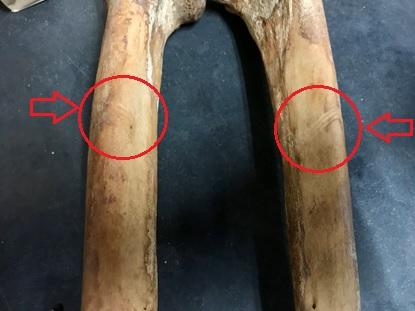
In the picture above, there are bilateral cut marks on the distal femora (the part of the thigh bones above the knee). As there are no signs of healing, the marks were determined to be peri- or postmortem. The next step is to establish if they occurred recently or historically. This is where staining can become very useful. If the damage occurred recently, the exposed bone would be white, contrasting with the surrounding cortical (outer) surface of the bone. While these marks are discolored, they nevertheless cut through and disrupt the previous staining pattern. Thus, we conclude that these are historic postmortem lacerations.
But how did they happen? We might never know for certain. During excavation, it was noted that the coffins were often buried in very close proximity, layered one on top of the other, or very close to each other. One possible explanation is that when a new grave was being dug, a tool such as a shovel passed through a coffin already in the ground, damaging the bones. This would explain the placement and symmetry of the marks. That being said, the cuts are not just a single line mirrored on the femora, but three parallel striations. Could a single blow from a shovel cause these marks? Or did the shovel hit the bones three separate times in nearly the same place? Or perhaps they came from a completely different source altogether. What do you think?
Reference:
Quatrehomme, Gérald, and Mehmet Yaşar Işcan. “Postmortem Skeletal Lesions.” Forensic Science International, vol. 89, no. 3, 1997, pp. 155–165.

